![]()
![]()
![]()
![]()
![]()
![]()
![]()
![]()
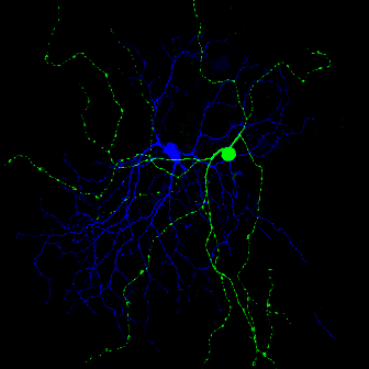
| Form and Function of Intrinsically Photosensitive Ganglion cells Another research program in our lab is to elucidate the structure and function of intrisically photosensitive ganglion cells in the mammalian retina. In mammals, photic information is exclusively processed by the retina and reaches the brain through the optic nerve. The eyes are equipped with at least two functionally and anatomically distinct light-detecting streams, the classic image-forming stream involving rods and cones and the non-image forming stream. The nonimage-forming photoreceptive stream entrains the circadian timing system and regulates pineal melatonin secretion and pupillary constriction. A small subpopulation of ganglion cells in the mammalian retina expresses the opsin-family photopigment melanopsin (Opn4). These ganglion cells are intrinsically photosensitive (ipRGC) and play a crucial role in non-image forming visual responses. Because melanopsin-containing ganglion cells are few in number and scattered throughout the retina, they are difficult to study. To address these limitations, we engineered a mouse line in which the Enhanced Green Fluorescent Protein (EGFP) is expressed under the control of the mouse melanopsin promoter employing BAC (bacterial artificial chromosome) transgenesis. We are performing two types of studies in these mice: (1) single cell electrophysiological recordings of EGFP-positive neurons in whole mount retinas to characterize their functional properties during development, (2) electrophysiological recordings of acutely isolated EGFP-positive neurons to characterize their intrinsic properties.
|