Spiking Initiated By Synaptic Inputs Using
The Five-Channel Model of Impulse Generation
|
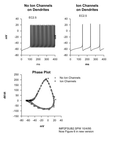
| Applying a current pulse to a retinal ganglion cell in a whole-cell recording mode generates impulse activity at a rate of about 1 impulse/pA/sec. A current pulse of several hundred ms evokes sustained firing from all types of ganglion cells, independent of how they respond to light (transient vs sustained, On vs Off, On-Off). In order to generate low frequency firing in computer simulations which attempt to model these results, the dendrites must have voltage-gated ion channels at lower density than that of the axon or soma; if not, the impulse firing goes to a much higher rate than that observed physiologically. This is illustrated in the two simulations (shown to the right) using and equivalent cylinder model without (upper left) and with (upper right) Na channels in the dendrites. The high rate of firing in the upper left panel can be attributed to passive dendrites which hold charge and continue to activate the impulse generation mechanism, whereas with active dendrites (upper right panel), the charge on the dendritic membrane is "wiped clean" by the active, antidromic impulse. |
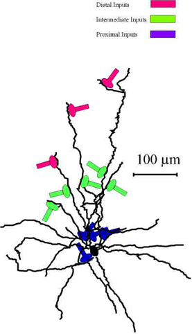
Simulations were made with realistic cell morphologies in which synaptic currents were generated via alpha-function conductance changes inserted at four different proximal dendritic sites (see the blue sites in the lefthand figure below). Each synaptic-like input (shown below, right) had a peak conductance of 2 nS, a peak delay of 50 ms and a reversal potential for the EPSP of 0 mV. The upper,
red trace, shows the timecourse of the membrane voltage (
EPSP in mV) for a single synaptic site. The lower,
blue trace, shows the timecourse of the voltage-clamped current (
EPSC) induced by the alpha-function conductance change for a single synaptic site.
References
Fohlmeister, J.F. and Miller, R.F. (1997) Impulse encoding mechanisms of ganglion cells in the tiger salamander retina. J. Neurophysiol. 78:1935-1947.
Fohlmeister, J.F. and Miller, R.F. (1997) Mechanisms by which cell geometry controls repetitive impulse firing in retinal ganglion cells. J. Neurophysiol. 78:1948-1964