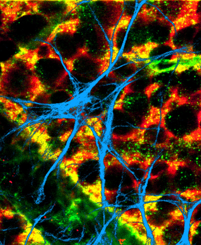
Astrocytes and Müller cells are shown in the following confocal immunofluorescence images of the vitreal surface of the rat retina. Astrocytes are labeled with antibodies against GFAP (glial fibrillary acid protein) and Müller cells with antibodies against vimentin or glutamine synthetase. Serial sections of confocal pictures were combined to form the images.
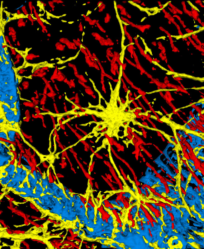
Astrocytes are shown in yellow, Müller cell endfeet in red, and blood vessels in blue in this triple-labeled fluorescence image. The three-dimensional surface rendering was created from a series of confocal images. The endfeet of the astrocytes and some of the Müller cells can be seen contacting blood vessels. Müller cells are labeled with antibodies against vimentin and blood vessels with antibodies against collagen IV. The width of the micrograph is 80 µm. (JPEG file, 316 KB.)
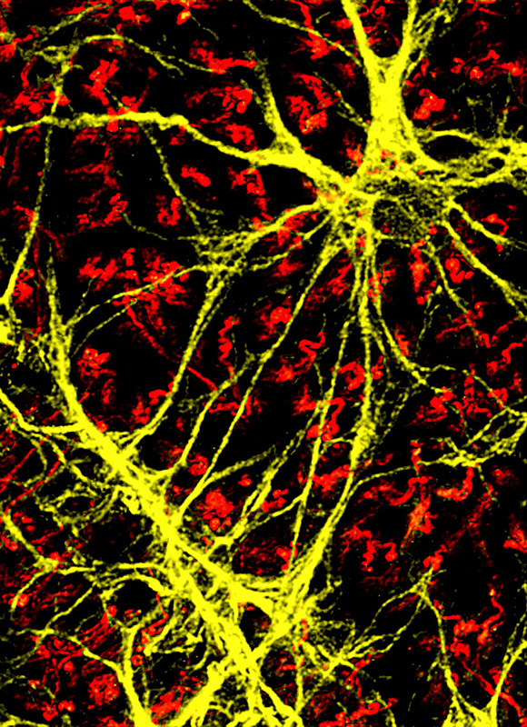
Astrocytes are shown in yellow and Müller cell endfeet in red in this double-labeled fluorescence image. Müller cells are labeled with antibodies against vimentin. The width of the micrograph is 75 µm. (JPEG file, 248 KB.)